
上海金畔生物科技有限公司代理AAT Bioquest荧光染料全线产品,欢迎访问AAT Bioquest荧光染料官网了解更多信息。
Stayright 紫色
 |
货号 | 45906 | 存储条件 | Multiple |
| 规格 | 50 mL | 价格 | 3924 | |
| Ex (nm) | Em (nm) | |||
| 分子量 | 1009.12 | 溶剂 | ||
| 产品详细介绍 | ||||
简要概述
3,3′-二氨基联苯胺(DAB)作为最常用的IHC色原已经应用了数十年,因为它便宜且对常规应用敏感。但是,已证明DAB具有诱变性,并且对实验室工作人员和环境有害。为了解决此问题,AAT提供的Stayright Purple是比DAB更安全的IHC色原。此外,Stayright Purple提供了一种快速简便的方法,可在HRP存在的情况下以DAB的高灵敏度产生干净而强烈的紫色荧光。 Stayright Purple HRP底物还显示出非诱变作用,细胞毒性最小。 ReadiUse Stayright 紫色过氧化物酶(HRP)底物适用于基于过氧化物酶(HRP)的免疫组织化学(IHC)和原位杂交(ISH)染色方法。在HRP诱导的氧化作用下,Stayright Purple在测定的目标部位形成紫色不溶性沉淀产物。最终的紫色物质不溶于有机溶剂和有机固定介质,因此可以通过常规的脱水和盖玻片步骤保持明显的紫色染料。为了提高便利性,您可以尝试使用我们的ReadiUse Stayright 紫色过氧化物酶(HRP)底物(#45900和45901)。它是一种稳定的,含有过氧化氢的预混合溶液,因此无需任何混合步骤,即可使用。金畔生物是AAT Bioquest的中国代理商,为您提供最优质的Stayright Purple IHC显色底物。
适用仪器
| 光学显微镜 | |
| 结构: | 白光 |
产品说明书
样品实验方案
简要概述
- 将有效的Stayright Purple溶液涂抹到组织切片上。
- 孵育组织切片5-15分钟。
- 漂洗组织5-10分钟,复染。
- 添加安装介质以覆盖该部分。
溶液配制
工作溶液配制
每1 mL Stayright Purple HRP缓冲液(组分B)中加入10μL100X Stayright Purple(组分A)和1μL稳定的3%过氧化氢(组分C)。
注意:未使用的预混合工作溶液可以在2-4℃下保存数周,以备将来使用。 但是,我们建议根据需要混合这些成分。
实验步骤
1.涂抹Stayright Purple溶液。 在室温下孵育5-15分钟。
2.将载玻片浸入dH2O中以停止颜色显影并检测染色强度。如果染色强度不够明亮,则需要更长的孵育时间。 您可以重新应用Stayright Purple溶液来继续开发。
3.用dH2O洗涤5-10分钟。
4.如果需要,请复染。
5.用乙醇脱水,然后永久固定在有机永久固定介质中。
图示
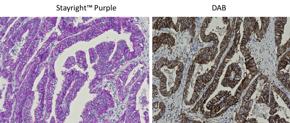 图1. FFPE肺腺癌组织中EpCAM的免疫组织化学检测。 将组织切片与多HRP共轭的山羊抗兔IgG孵育,然后分别用Stayright Purple(左)和DAB(右)显影。 细胞用苏木精复染。 Stayright Purple产生的深层荧光具有高灵敏度和清晰的分辨率,类似于DAB。 |
参考文献
3,3′-diaminobenzidine (DAB)-H2O2-HRP voltammetric enzyme-linked immunoassay for the detection of carcionembryonic antigen
Authors: Zhang, S., Yang, J., Lin, J.
Journal: Bioelectrochemistry (2008): 47-52
HistoGreen: a new alternative to 3,3′-diaminobenzidine-tetrahydrochloride-dihydrate (DAB) as a peroxidase substrate in immunohistochemistry?
Authors: Thomas, M. A., Lemmer, B.
Journal: Brain Res Brain Res Protoc (2005): 107-18
Stability and solubility of 3,3′-diaminobenzidine (DAB)
Authors: Kiernan, J. A.
Journal: Biotech Histochem (2003): 135
Safe diaminobenzidine (DAB) disposal
Authors: Horn, H.
Journal: Biotech Histochem (2002): 229
Non-fluorescent chromosome painting using the peroxidase/diaminobenzidine (DAB) reaction
Authors: K and a, R., Suzuki, M., Minamihisamatsu, M., Furukawa, A., Odaka, T., Hayata, I.
Journal: Int J Radiat Biol (1998): 529-33
Modified cerium-based and Gomori-based cerium methods for light microscopic phosphatase histochemistry: the cerium-perhydroxide-diaminobenzidine-nickel (Ce-H2O2-DAB-Ni and Ce/Ce-H2O2-DAB-Ni) two-step procedures
Authors: Halbhuber, K. J., Feuerstein, H., Moller, U., Klinger, M.
Journal: Acta Histochem (1992): 87-103
Employment of merocyanine 540 fluorescence to form diaminobenzidine (DAB) oxidation product: a photoconversion method for the visualization of erythrocyte membrane fluidity for light and electron microscopy
Authors: Oehring, H., Halbhuber, K. J.
Journal: Acta Histochem (1991): 127-34
Copper-H2O2 oxidation strikingly improves silver intensification of the nickel-diaminobenzidine (Ni-DAB) end-product of the peroxidase reaction
Authors: Gallyas, F., Merchenthaler, I.
Journal: J Histochem Cytochem (1988): 807-10
The cerium perhydroxide-diaminobenzidine (Ce-H2O2-DAB) procedure. New methods for light microscopic phosphatase histochemistry and immunohistochemistry
Authors: Halbhuber, K. J., Gossrau, R., Moller, U., Hulstaert, C. E., Zimmermann, N., Feuerstein, H.
Journal: Histochemistry (1988): 289-97
The use of gold-substituted silver-intensified diaminobenzidine (DAB) and non-intensified DAB for simultaneous electron microscopic immunoperoxidase labeling of tyrosine hydroxylase and glutamic acid decarboxylase immunoreactivity in the rat medial preoptic area
Authors: Gorcs, T. J., Leranth, C., MacLusky, N. J.
Journal: J Histochem Cytochem (1986): 1439-47