钙离子荧光探针Rhod-4, AM 货号21123-AAT Bioquest荧光染料

上海金畔生物科技有限公司代理AAT Bioquest荧光染料全线产品,欢迎访问AAT Bioquest荧光染料官网了解更多信息。

钙离子荧光探针Rhod-4, AM

钙离子荧光探针Rhod-4, AM

钙离子荧光探针Rhod-4, AM    货号21123 货号 21123 存储条件 在零下15度以下保存, 避免光照
规格 20×50 ug 价格 6564
Ex (nm) 523 Em (nm) 551
分子量 1015.96 溶剂 DMSO
产品详细介绍

简要概述

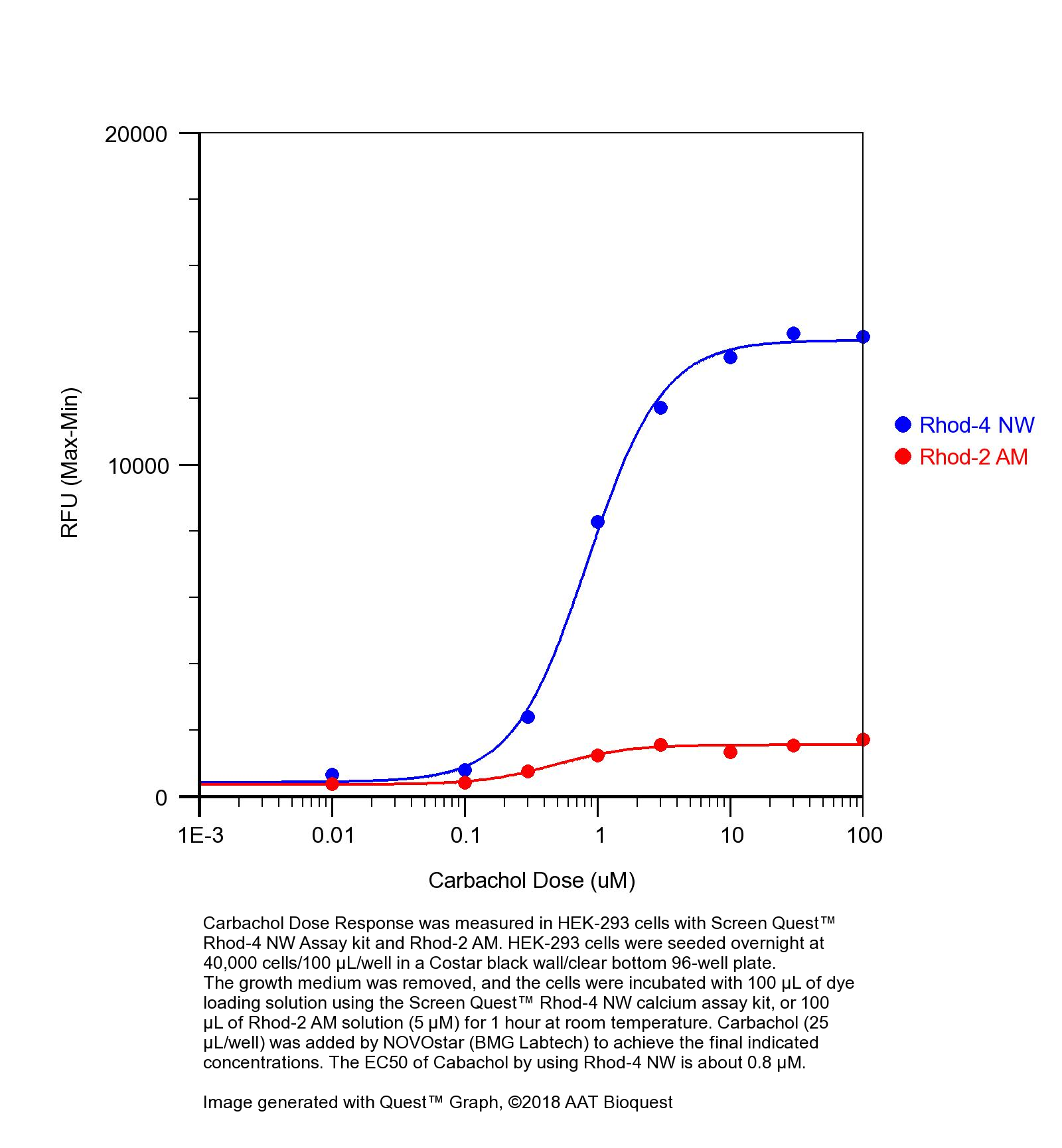
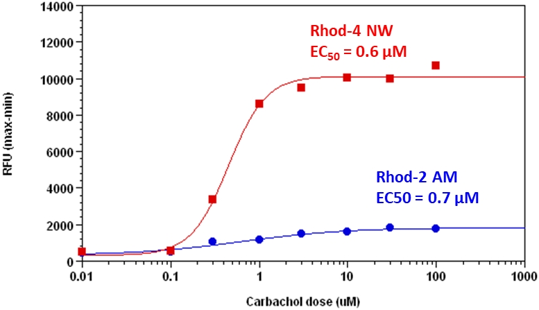
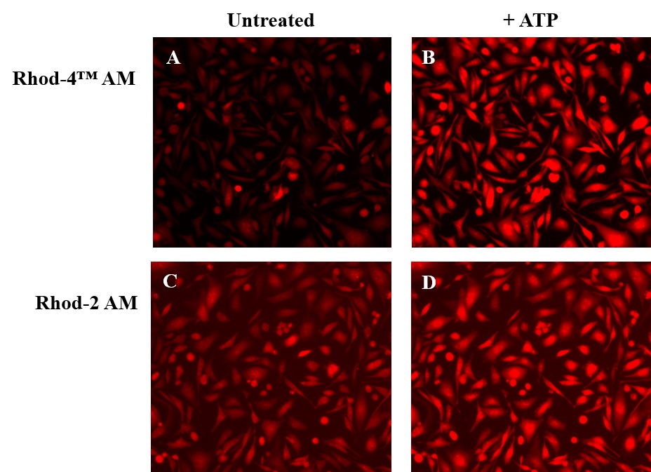
钙离子荧光探针Rhod-4, AM是美国AAT Bioquest生产的用于标记钙离子的荧光探针,钙的测量对于许多生物学研究至关重要。荧光探针显示结合Ca2+后的光谱响应,使研究人员能够通过荧光显微镜,流式细胞仪,荧光光谱和荧光酶标仪研究细胞内游离Ca2+浓度的变化。Rhod-2最常用于红色荧光钙指示剂中。然而,Rhod-2 AM在酯酶水解后仅在活细胞中发生中度荧光,并且具有非常小的细胞钙响应。Rhod-4 已被开发用于改善Rhod-2细胞负载和钙响应,同时保持Rhod-2的光谱波长。在CHO和HEK细胞中,Rhod-4 AM具有比Rhod-2 AM敏感10倍的细胞钙响应。AAT Bioquest提供多种包装尺寸的Quest Rhod-4,以满足您的特殊需求,例如1毫克; 10×50μg; 20×50μg; HTS包装,无需额外包装费用。金畔生物是AAT Bioquest 的中国代理商,为您提供最优质的钙离子荧光探针。

点击查看光谱

点击查看实验方案

钙离子篇:时间轴式讲解应用于钙离子检测的探针


适用仪器


荧光显微镜  
激发: TRITC
发射: TRITC
推荐孔板: 黑色透明
荧光酶标仪  
激发: 540nm
发射: 590nm
cutoff: 570nm
推荐孔板: 黑色透明
读取模式: 底读模式

产品说明书

操作步骤

1.准备HHBS缓冲液,10%Pluronic®F-127溶液和25 mM Probenecid溶液。
 

2.在高质量无水DMSO中制备2 mM至5 mM Rhod-4 ,AM原液。
2.1使用量的Rhod-4 ,AM:1 mg
2.2所需浓度:2 mM
2.3在合适的容器中,将1mg Rhod-4,AM与492.15μL无水DMSO混合。
 

3.使用10μMRhod-4 ,AM 4在HHBS中制备2X工作溶液,0.08%Pluronic®F-127和2 mM丙磺舒。
3.1最终井内浓度的Rhod-4 ,AM:5μM
3.2Pluronic®F-127的最终井内浓度:0.04%
3.3最终孔内浓度的丙磺舒:1mM
3.4在合适的容器中混合16μL的Rhod-4 ,AM,25.6μL的10%Pluronic F-127和256μL的25mM丙磺舒。然后,添加HHBS或您选择的缓冲液,直到体积为3.2 mL。
注意:对于大多数细胞系,我们建议使用Rhod-4 的最终浓度,AM为4至5μM。
注意:推荐的Pluronic F-127孔浓度最终为0.02%至0.04%。
注意:推荐的最终浓度为1至2.5 mM的Probenecid。
 

4.将100μL染料工作溶液加入已经含有100μL培养基的所需孔中。
4.1该步骤将染料工作溶液从2X稀释至1X,并将每种组分的最终浓度调节至以下:5μM的Rhod-4 ,AM,0.04%Pluronic F-127,1mM丙磺舒。
 

5.孵育染料
5.1将染料加载板在细胞培养箱中孵育20-120分钟。
5.2将染料加载板在室温下孵育30分钟。
 

6.用1.0 mM Probenecid准备HHBS缓冲液(或您选择的缓冲液)。
6.1在合适的容器中加入160μL的25mM丙磺舒。接下来,添加HHBS或您选择的缓冲液,直到体积为4 mL。
 

7.用HHBS缓冲液或您选择的缓冲液替换染料工作溶液,使用1.0 mM Probenecid。
7.1首先,从所需孔中除去200μL染料工作溶液和培养基。
7.2在相同的孔中加入200μL含有1.0mM丙磺舒的HHBS(或您选择的缓冲液)。
 

8.运行实验
8.1为您的样品添加所需的处理。
8.2以Ex / Em = 524/551 nm运行实验。


参考文献

Central role of IP 3 R2-mediated Ca 2+ oscillation in self-renewal of liver cancer stem cells elucidated by high-signal ER sensor
Authors: Cuiwei Sun, Bo Shui, Wei Zhao, Hui Liu, Wenwen Li, Jane C Lee, Robert Doran, Frank K Lee, Tao Sun, Qing Sunny Shen
Journal: Cell death & disease (2019): 396

Imaging elemental events of store-operated Ca2+ entry in invading cancer cells with plasmalemmal targeted sensors
Authors: Fujian Lu, Jianwei Sun, Qiaoxia Zheng, Jinghang Li, Yuanzhao Hu, Peng Yu, Huifang He, Yan Zhao, Xianhua Wang, Shengyu Yang
Journal: J Cell Sci (2019): jcs–224923

Autocrine GABA signaling distinctively regulates phenotypic activation of mouse pulmonary macrophages
Authors: Luan Januzi, Jacob W Poirier, Matthew JE Maksoud, Yun-Yan Xiang, Rudolf AW Veldhuizen, Sean E Gill, Sean P Cregan, Haibo Zhang, Gregory A Dekaban, Wei-Yang Lu
Journal: Cellular Immunology (2018)

Three-dimensional model of intracellular and intercellular Ca2+ waves propagation in endothelial cells
Authors: Toshihiro Sera, Shingo Komine, Masataka Arai, Yasuhiro Sunaga, Hideo Yokota, Susumu Kudo
Journal: Biochemical and Biophysical Research Communications (2018)

Effect of stem cell niche elasticity/ECM protein on the self-beating cardiomyocyte differentiation of inducedpluripotent stem (iPS) cells at different stages
Authors: Mitsuhi Hirata, Tetsuji Yamaoka
Journal: Acta Biomaterialia (2017)

Emerin plays a crucial role in nuclear invagination and in the nuclear calcium transient
Authors: Masaya Shimojima, Shinsuke Yuasa, Chikaaki Motoda, Gakuto Yozu, Toshihiro Nagai, Shogo Ito, Mark Lachmann, Shin Kashimura, Makoto Takei, Dai Kusumoto
Journal: Scientific Reports (2017)

Preliminary findings on ultrasound modulation of the electromechanical function of human stem-cell-derived cardiomyocytes
Authors: Andrew William Chen, Aleksandra Klimas, Vesna Zderic, Ivan Suares Castellanos, Emilia Entcheva
Journal: (2017): 1–4

The role of spatial organization of Ca (2+) release sites in the generation of arrhythmogenic diastolic Ca (2+) release in myocytes from failing hearts.
Authors: Andriy E Belevych, Hsiang-Ting Ho, Ingrid M Bonilla, Radmila Terentyeva, Karsten E Schober, Dmitry Terentyev, Cynthia A Carnes, Sándor Györke
Journal: Basic research in cardiology (2017): 44

Dynamic polyrotaxane-coated surface for effective differentiation of mouse induced pluripotent stem cells into cardiomyocytes
Authors: Ji-Hun Seo, Mitsuhi Hirata, Sachiro Kakinoki, Tetsuji Yamaoka, Nobuhiko Yui
Journal: RSC Advances (2016): 35668–35676

Individual evaluation of cardiac marker expression and self-beating during cardiac differentiation of P19CL6 cells on different culture substrates
Authors: Tetsuji Yamaoka, Mitsuhi Hirata, Takaaki Dan, Atsushi Yamashita, Akihisa Otaka, Takahiko Nakaoki, Azizi Miskon, Sachiro Kakinoki, Atsushi Mahara
Journal: Journal of Biomedical Materials Research Part A (2016)

说明书
钙离子荧光探针Rhod-4, AM.pdf

Screen Quest 免洗Rhod-4钙检测试剂盒 去除培养基 货号36330-AAT Bioquest荧光染料

上海金畔生物科技有限公司代理AAT Bioquest荧光染料全线产品,欢迎访问AAT Bioquest荧光染料官网了解更多信息。

Screen Quest 免洗Rhod-4钙检测试剂盒 去除培养基

Screen Quest 免洗Rhod-4钙检测试剂盒 去除培养基

货号 36330 存储条件 在零下15度以下保存, 避免光照
规格 1 Plate 价格 1272
Ex (nm) 523 Em (nm) 551
分子量 溶剂
产品详细介绍

简要概述

钙通量测定法是药物发现中筛选G蛋白偶联受体(GPCR)的首选方法。 Screen Quest Rhod-4 NW钙含量测定试剂盒提供了一种基于荧光的均相测定,用于检测细胞内钙的迁移。Rhod-4是可用于HTS筛选的最亮的红色钙指示剂。一旦进入细胞内,Rhod-4 的亲脂性封闭基团就会被非特异性细胞酯酶裂解,产生带负电荷的荧光染料,并留在细胞内部,并且与钙结合后其荧光会大大增强。当细胞被筛选化合物刺激时,受体信号释放细胞内钙,这大大增加了Rhod-4的荧光。 Rhod-4 具有长波长,高灵敏度和大于250倍的荧光增强特性(当与钙形成复合物时),使其成为测量细胞钙的理想指标。此Screen Quest Rhod-4 NW钙测定试剂盒提供了一种优化的测定方法,用于检测G蛋白偶联受体(GPCR)和钙通道。该测定可以用96孔或384孔微孔板进行,并易于适应自动化。金畔生物是AAT Bioquest的中国代理商,为您提供最优质的Screen Quest 免洗Rhod-4钙检测试剂盒。

点击查看光谱


适用仪器


荧光酶标仪  
激发: 540nm
发射: 590nm
cutoff: 570nm
推荐孔板: 黑色透明
读取模式: 底读模式
其他可用仪器:
FDSS, FLIPR, ViewLux, NOVOStar, ArrayScan, FlexStation, IN Cell Analyzer

产品说明书

样品实验方案

简要概述

  1. 准备细胞
  2. 除去生长培养基
  3. 添加Rhod-4 NW染料加载溶液(对于96孔板为100 µL /孔,对于384孔板为25 µL /孔)
  4. 在室温下孵育1小时
  5. 检测Ex / Em = 540/590 nm的荧光强度


溶液配制

储备溶液配制

1. Rhod-4 NW储备溶液:将100 µL DMSO加入小瓶Rhod-4 NW(组分A)中并充分混合,避光。 注意:10 µL Rhod-4 NW储备液足以装满一块板。

2.分析缓冲液(1X):a)对于#36330(1个板试剂盒)和#363331(10个板试剂盒),通过将9 mL HHBS(组分C)添加到10XPluronic®F127 Plus(1 mL,组分B)中来制成1X分析缓冲液,并充分混合。b)对于#36332(100板试剂盒),通过向10XPluronic®F127 Plus(10 mL,组分B)中加入90 mL HHBS(未包括)制成1X Assay Buffer。 注意:10 mL 1X分析缓冲液足以用于一块板。 分装并在<-20℃下存储未使用的1X分析缓冲液。 避光并避免重复的冻融循环。


工作溶液配制

Rhod-4 NW上色溶液:将10 µL Rhod-4 NW储备液添加到10 mL 1X分析缓冲液中,并充分混合。 注意:该工作溶液在室温下至少可稳定2小时。


实验步骤

1.从细胞板中去除生长培养基。注意:重要的是去除生长培养基,以最大程度地减少背景荧光和化合物对血清或培养基的干扰。或者,使细胞在含1-5%FBS的生长培养基中生长,以避免培养基去除步骤。在这种情况下,需要在HHBS缓冲液中添加2X染料。 [我们为在生长培养基中使用0.5-1%FBS以避免培养基去除步骤的人提供2个单独的免洗钙测定试剂盒(Cat.#36334和Cat.#36335)。

2.将100 µL /孔(96孔板)或25 µL /孔(384孔板)的Rhod-4 NW染料加载溶液添加到细胞板中。

3.在细胞培养箱中将染料加载板孵育30分钟,然后在室温下再孵育30分钟。注意:如果测定需要37°C,请立即进行实验,而无需进一步室温孵育。如果细胞在室温下能长时间正常工作,则在室温下孵育细胞板1-2小时。

4.用HHBS或所需的缓冲液准备复合板。

5.通过检测Ex / Em = 540/590 nm的荧光强度进行钙通量检测。


图示

Screen Quest 免洗Rhod-4钙检测试剂盒 去除培养基   货号36330

图1.使用Screen Quest Rhod-4 NW分析试剂盒和Rhod-2 AM在HEK-293细胞中测量了卡巴胆碱剂量反应。 将HEK-293细胞以40,000个细胞/ 100 µL /孔在Costar黑色96孔板中过夜接种。 去除生长培养基,并使用Screen Quest Rhod-4 NW钙分析试剂盒将细胞与100 µL染料上样溶液或室温下100 µL Rhod-2 AM溶液(5 µM)孵育1小时。 NOVOstar(BMG Labtech)添加了卡巴胆碱(25 µL /孔)以达到最终指示的浓度。 使用Rhod-4 NW时卡巴胆的EC50约为0.8 µM。


参考文献

Fluorescence absorbance inner-filter decomposition: the role of emission shape on estimates of free Ca(2+) using Rhod-2
Authors: Territo PR, Heil J, Bose S, Evans FJ, Balaban RS.
Journal: Appl Spectrosc (2007): 138

Kinetic characterization of novel NR2B antagonists using fluorescence detection of calcium flux
Authors: Bednar B, Cunningham ME, Kiss L, Cheng G, McCauley JA, Liverton NJ, Koblan KS.
Journal: J Neurosci Methods (2004): 247

Novel fluo-4 analogs for fluorescent calcium measurements
Authors: Martin VV, Beierlein M, Morgan JL, Rothe A, Gee KR.
Journal: Cell Calcium (2004): 509

Protein kinase C and myocardial calcium handling during ischemia and reperfusion: lessons learned using Rhod-2 spectrofluorometry
Authors: Stamm C, del Nido PJ.
Journal: Thorac Cardiovasc Surg (2004): 127

Cytosolic calcium in the ischemic rabbit heart: assessment by pH- and temperature-adjusted rhod-2 spectrofluorometry
Authors: Stamm C, Friehs I, Choi YH, Zurakowski D, McGowan FX, del Nido PJ.
Journal: Cardiovasc Res (2003): 695

Calcium measurements in perfused mouse heart: quantitating fluorescence and absorbance of Rhod-2 by application of photon migration theory
Authors: Du C, MacGowan GA, Farkas DL, Koretsky AP.
Journal: Biophys J (2001): 549

Calibration of the calcium dissociation constant of Rhod(2)in the perfused mouse heart using manganese quenching
Authors: Du C, MacGowan GA, Farkas DL, Koretsky AP.
Journal: Cell Calcium (2001): 217

Changes in mitochondrial Ca2+ detected with Rhod-2 in single frog and mouse skeletal muscle fibres during and after repeated tetanic contractions
Authors: Lannergren J, Westerblad H, Bruton JD.
Journal: J Muscle Res Cell Motil (2001): 265

Rhod-2 based measurements of intracellular calcium in the perfused mouse heart: cellular and subcellular localization and response to positive inotropy
Authors: MacGowan GA, Du C, Glonty V, Suhan JP, Koretsky AP, Farkas DL.
Journal: J Biomed Opt (2001): 23

Mitochondrial free calcium levels (Rhod-2 fluorescence) and ultrastructural alterations in neuronally differentiated PC12 cells during ceramide-dependent cell death
Authors: Muriel MP, Lambeng N, Darios F, Michel PP, Hirsch EC, Agid Y, Ruberg M.
Journal: J Comp Neurol (2000): 297

说明书
Screen Quest 免洗Rhod-4钙检测试剂盒 去除培养基.pdf

Screen Quest 免洗Rhod-4钙检测试剂盒 去除培养基 10板 货号36331-AAT Bioquest荧光染料

上海金畔生物科技有限公司代理AAT Bioquest荧光染料全线产品,欢迎访问AAT Bioquest荧光染料官网了解更多信息。

Screen Quest 免洗Rhod-4钙检测试剂盒 去除培养基 10板

Screen Quest 免洗Rhod-4钙检测试剂盒 去除培养基 10板

货号 36331 存储条件 在零下15度以下保存, 避免光照
规格 10 Plates 价格 5244
Ex (nm) 523 Em (nm) 551
分子量 溶剂
产品详细介绍

简要概述

钙通量测定法是药物发现中筛选G蛋白偶联受体(GPCR)的首选方法。 Screen Quest Rhod-4 NW钙含量测定试剂盒提供了一种基于荧光的均相测定,用于检测细胞内钙的迁移。Rhod-4是可用于HTS筛选的最亮的红色钙指示剂。一旦进入细胞内,Rhod-4 的亲脂性封闭基团就会被非特异性细胞酯酶裂解,产生带负电荷的荧光染料,并留在细胞内部,并且与钙结合后其荧光会大大增强。当细胞被筛选化合物刺激时,受体信号释放细胞内钙,这大大增加了Rhod-4的荧光。 Rhod-4 具有长波长,高灵敏度和大于250倍的荧光增强特性(当与钙形成复合物时),使其成为测量细胞钙的理想指标。此Screen Quest Rhod-4 NW钙测定试剂盒提供了一种优化的测定方法,用于检测G蛋白偶联受体(GPCR)和钙通道。该测定可以用96孔或384孔微孔板进行,并易于适应自动化。金畔生物是AAT Bioquest的中国代理商,为您提供最优质的Screen Quest 免洗Rhod-4钙检测试剂盒。

点击查看光谱


适用仪器


荧光酶标仪  
激发: 540nm
发射: 590nm
cutoff: 570nm
推荐孔板: 黑色透明
读取模式: 底读模式
其他可用仪器:
FDSS, FLIPR, ViewLux, NOVOStar, ArrayScan, FlexStation, IN Cell Analyzer

产品说明书

样品实验方案

简要概述

  1. 准备细胞
  2. 除去生长培养基
  3. 添加Rhod-4 NW染料加载溶液(对于96孔板为100 µL /孔,对于384孔板为25 µL /孔)
  4. 在室温下孵育1小时
  5. 检测Ex / Em = 540/590 nm的荧光强度


溶液配制

储备溶液配制

1. Rhod-4 NW储备溶液:将100 µL DMSO加入小瓶Rhod-4 NW(组分A)中并充分混合,避光。 注意:10 µL Rhod-4 NW储备液足以装满一块板。

2.分析缓冲液(1X):a)对于#36330(1个板试剂盒)和#363331(10个板试剂盒),通过将9 mL HHBS(组分C)添加到10XPluronic®F127 Plus(1 mL,组分B)中来制成1X分析缓冲液,并充分混合。b)对于#36332(100板试剂盒),通过向10XPluronic®F127 Plus(10 mL,组分B)中加入90 mL HHBS(未包括)制成1X Assay Buffer。 注意:10 mL 1X分析缓冲液足以用于一块板。 分装并在<-20℃下存储未使用的1X分析缓冲液。 避光并避免重复的冻融循环。


工作溶液配制

Rhod-4 NW上色溶液:将10 µL Rhod-4 NW储备液添加到10 mL 1X分析缓冲液中,并充分混合。 注意:该工作溶液在室温下至少可稳定2小时。


实验步骤

1.从细胞板中去除生长培养基。注意:重要的是去除生长培养基,以最大程度地减少背景荧光和化合物对血清或培养基的干扰。或者,使细胞在含1-5%FBS的生长培养基中生长,以避免培养基去除步骤。在这种情况下,需要在HHBS缓冲液中添加2X染料。 [我们为在生长培养基中使用0.5-1%FBS以避免培养基去除步骤的人提供2个单独的免洗钙测定试剂盒(Cat.#36334和Cat.#36335)。

2.将100 µL /孔(96孔板)或25 µL /孔(384孔板)的Rhod-4 NW染料加载溶液添加到细胞板中。

3.在细胞培养箱中将染料加载板孵育30分钟,然后在室温下再孵育30分钟。注意:如果测定需要37°C,请立即进行实验,而无需进一步室温孵育。如果细胞在室温下能长时间正常工作,则在室温下孵育细胞板1-2小时。

4.用HHBS或所需的缓冲液准备复合板。

5.通过检测Ex / Em = 540/590 nm的荧光强度进行钙通量检测。


图示

Screen Quest 免洗Rhod-4钙检测试剂盒 去除培养基 10板   货号36331

图1.使用Screen Quest Rhod-4 NW分析试剂盒和Rhod-2 AM在HEK-293细胞中测量了卡巴胆碱剂量反应。 将HEK-293细胞以40,000个细胞/ 100 µL /孔在Costar黑色96孔板中过夜接种。 去除生长培养基,并使用Screen Quest Rhod-4 NW钙分析试剂盒将细胞与100 µL染料上样溶液或室温下100 µL Rhod-2 AM溶液(5 µM)孵育1小时。 NOVOstar(BMG Labtech)添加了卡巴胆碱(25 µL /孔)以达到最终指示的浓度。 使用Rhod-4 NW时卡巴胆的EC50约为0.8 µM。


参考文献

Fluorescence absorbance inner-filter decomposition: the role of emission shape on estimates of free Ca(2+) using Rhod-2
Authors: Territo PR, Heil J, Bose S, Evans FJ, Balaban RS.
Journal: Appl Spectrosc (2007): 138

Kinetic characterization of novel NR2B antagonists using fluorescence detection of calcium flux
Authors: Bednar B, Cunningham ME, Kiss L, Cheng G, McCauley JA, Liverton NJ, Koblan KS.
Journal: J Neurosci Methods (2004): 247

Novel fluo-4 analogs for fluorescent calcium measurements
Authors: Martin VV, Beierlein M, Morgan JL, Rothe A, Gee KR.
Journal: Cell Calcium (2004): 509

Protein kinase C and myocardial calcium handling during ischemia and reperfusion: lessons learned using Rhod-2 spectrofluorometry
Authors: Stamm C, del Nido PJ.
Journal: Thorac Cardiovasc Surg (2004): 127

Cytosolic calcium in the ischemic rabbit heart: assessment by pH- and temperature-adjusted rhod-2 spectrofluorometry
Authors: Stamm C, Friehs I, Choi YH, Zurakowski D, McGowan FX, del Nido PJ.
Journal: Cardiovasc Res (2003): 695

Calcium measurements in perfused mouse heart: quantitating fluorescence and absorbance of Rhod-2 by application of photon migration theory
Authors: Du C, MacGowan GA, Farkas DL, Koretsky AP.
Journal: Biophys J (2001): 549

Calibration of the calcium dissociation constant of Rhod(2)in the perfused mouse heart using manganese quenching
Authors: Du C, MacGowan GA, Farkas DL, Koretsky AP.
Journal: Cell Calcium (2001): 217

Changes in mitochondrial Ca2+ detected with Rhod-2 in single frog and mouse skeletal muscle fibres during and after repeated tetanic contractions
Authors: Lannergren J, Westerblad H, Bruton JD.
Journal: J Muscle Res Cell Motil (2001): 265

Rhod-2 based measurements of intracellular calcium in the perfused mouse heart: cellular and subcellular localization and response to positive inotropy
Authors: MacGowan GA, Du C, Glonty V, Suhan JP, Koretsky AP, Farkas DL.
Journal: J Biomed Opt (2001): 23

Mitochondrial free calcium levels (Rhod-2 fluorescence) and ultrastructural alterations in neuronally differentiated PC12 cells during ceramide-dependent cell death
Authors: Muriel MP, Lambeng N, Darios F, Michel PP, Hirsch EC, Agid Y, Ruberg M.
Journal: J Comp Neurol (2000): 297

说明书
Screen Quest 免洗Rhod-4钙检测试剂盒 去除培养基 10板.pdf

Screen Quest 免洗Rhod-4钙检测试剂盒 去除培养基 10×10板 货号36332-AAT Bioquest荧光染料

上海金畔生物科技有限公司代理AAT Bioquest荧光染料全线产品,欢迎访问AAT Bioquest荧光染料官网了解更多信息。

Screen Quest 免洗Rhod-4钙检测试剂盒 去除培养基 10×10板

Screen Quest 免洗Rhod-4钙检测试剂盒 去除培养基 10×10板

货号 36332 存储条件 在零下15度以下保存, 避免光照
规格 100 Plates 价格 39060
Ex (nm) 523 Em (nm) 551
分子量 溶剂
产品详细介绍

简要概述

钙通量测定法是药物发现中筛选G蛋白偶联受体(GPCR)的首选方法。 Screen Quest Rhod-4 NW钙含量测定试剂盒提供了一种基于荧光的均相测定,用于检测细胞内钙的迁移。Rhod-4是可用于HTS筛选的最亮的红色钙指示剂。一旦进入细胞内,Rhod-4 的亲脂性封闭基团就会被非特异性细胞酯酶裂解,产生带负电荷的荧光染料,并留在细胞内部,并且与钙结合后其荧光会大大增强。当细胞被筛选化合物刺激时,受体信号释放细胞内钙,这大大增加了Rhod-4的荧光。 Rhod-4 具有长波长,高灵敏度和大于250倍的荧光增强特性(当与钙形成复合物时),使其成为测量细胞钙的理想指标。此Screen Quest Rhod-4 NW钙测定试剂盒提供了一种优化的测定方法,用于检测G蛋白偶联受体(GPCR)和钙通道。该测定可以用96孔或384孔微孔板进行,并易于适应自动化。金畔生物是AAT Bioquest的中国代理商,为您提供最优质的Screen Quest 免洗Rhod-4钙检测试剂盒。

点击查看光谱


适用仪器


荧光酶标仪  
激发: 540nm
发射: 590nm
cutoff: 570nm
推荐孔板: 黑色透明
读取模式: 底读模式
其他可用仪器:
FDSS, FLIPR, ViewLux, NOVOStar, ArrayScan, FlexStation, IN Cell Analyzer

产品说明书

样品实验方案

简要概述

  1. 准备细胞
  2. 除去生长培养基
  3. 添加Rhod-4 NW染料加载溶液(对于96孔板为100 µL /孔,对于384孔板为25 µL /孔)
  4. 在室温下孵育1小时
  5. 检测Ex / Em = 540/590 nm的荧光强度


溶液配制

储备溶液配制

1. Rhod-4 NW储备溶液:将100 µL DMSO加入小瓶Rhod-4 NW(组分A)中并充分混合,避光。 注意:10 µL Rhod-4 NW储备液足以装满一块板。

2.分析缓冲液(1X):a)对于#36330(1个板试剂盒)和#363331(10个板试剂盒),通过将9 mL HHBS(组分C)添加到10XPluronic®F127 Plus(1 mL,组分B)中来制成1X分析缓冲液,并充分混合。b)对于#36332(100板试剂盒),通过向10XPluronic®F127 Plus(10 mL,组分B)中加入90 mL HHBS(未包括)制成1X Assay Buffer。 注意:10 mL 1X分析缓冲液足以用于一块板。 分装并在<-20℃下存储未使用的1X分析缓冲液。 避光并避免重复的冻融循环。


工作溶液配制

Rhod-4 NW工作溶液:将20 µL Rhod-4 NW储备溶液加入10 mL 1X分析缓冲液中,并充分混合。 注意:该工作溶液在室温下至少可稳定2小时。


实验步骤

1.将100 µL /孔(96孔板)或25 µL /孔(384孔板)的Rhod-4 NW染料加载溶液添加到细胞板中。

2.在细胞培养箱中将染料加载板孵育30分钟,然后在室温下再孵育30分钟。 注意:如果测定需要37℃,请立即进行实验,而无需进一步室温孵育。 注意:如果细胞在室温下能长时间正常工作,请在室温下孵育细胞板1-2小时。
 
3.准备并添加HHBS或所需缓冲液的复合板。

4.通过检测Ex / Em = 540/590 nm的荧光强度运行钙通量检测。


图示

Screen Quest 免洗Rhod-4钙检测试剂盒 去除培养基 10x10板    货号36332

图1.使用Screen Quest Rhod-4 NW分析试剂盒和Rhod-2 AM在HEK-293细胞中测量了卡巴胆碱剂量反应。 将HEK-293细胞以40,000个细胞/ 100 µL /孔在Costar黑色96孔板中过夜接种。 去除生长培养基,并使用Screen Quest Rhod-4 NW钙分析试剂盒将细胞与100 µL染料上样溶液或室温下100 µL Rhod-2 AM溶液(5 µM)孵育1小时。 NOVOstar(BMG Labtech)添加了卡巴胆碱(25 µL /孔)以达到最终指示的浓度。 使用Rhod-4 NW时卡巴胆的EC50约为0.8 µM。


参考文献

Fluorescence absorbance inner-filter decomposition: the role of emission shape on estimates of free Ca(2+) using Rhod-2
Authors: Territo PR, Heil J, Bose S, Evans FJ, Balaban RS.
Journal: Appl Spectrosc (2007): 138

Kinetic characterization of novel NR2B antagonists using fluorescence detection of calcium flux
Authors: Bednar B, Cunningham ME, Kiss L, Cheng G, McCauley JA, Liverton NJ, Koblan KS.
Journal: J Neurosci Methods (2004): 247

Novel fluo-4 analogs for fluorescent calcium measurements
Authors: Martin VV, Beierlein M, Morgan JL, Rothe A, Gee KR.
Journal: Cell Calcium (2004): 509

Protein kinase C and myocardial calcium handling during ischemia and reperfusion: lessons learned using Rhod-2 spectrofluorometry
Authors: Stamm C, del Nido PJ.
Journal: Thorac Cardiovasc Surg (2004): 127

Cytosolic calcium in the ischemic rabbit heart: assessment by pH- and temperature-adjusted rhod-2 spectrofluorometry
Authors: Stamm C, Friehs I, Choi YH, Zurakowski D, McGowan FX, del Nido PJ.
Journal: Cardiovasc Res (2003): 695

Calcium measurements in perfused mouse heart: quantitating fluorescence and absorbance of Rhod-2 by application of photon migration theory
Authors: Du C, MacGowan GA, Farkas DL, Koretsky AP.
Journal: Biophys J (2001): 549

Calibration of the calcium dissociation constant of Rhod(2)in the perfused mouse heart using manganese quenching
Authors: Du C, MacGowan GA, Farkas DL, Koretsky AP.
Journal: Cell Calcium (2001): 217

Changes in mitochondrial Ca2+ detected with Rhod-2 in single frog and mouse skeletal muscle fibres during and after repeated tetanic contractions
Authors: Lannergren J, Westerblad H, Bruton JD.
Journal: J Muscle Res Cell Motil (2001): 265

Rhod-2 based measurements of intracellular calcium in the perfused mouse heart: cellular and subcellular localization and response to positive inotropy
Authors: MacGowan GA, Du C, Glonty V, Suhan JP, Koretsky AP, Farkas DL.
Journal: J Biomed Opt (2001): 23

Mitochondrial free calcium levels (Rhod-2 fluorescence) and ultrastructural alterations in neuronally differentiated PC12 cells during ceramide-dependent cell death
Authors: Muriel MP, Lambeng N, Darios F, Michel PP, Hirsch EC, Agid Y, Ruberg M.
Journal: J Comp Neurol (2000): 297

说明书
Screen Quest 免洗Rhod-4钙检测试剂盒 去除培养基 10×10板 .pdf

Screen Quest 免洗Rhod-4钙检测试剂盒 货号36333-AAT Bioquest荧光染料

上海金畔生物科技有限公司代理AAT Bioquest荧光染料全线产品,欢迎访问AAT Bioquest荧光染料官网了解更多信息。

Screen Quest 免洗Rhod-4钙检测试剂盒

Screen Quest 免洗Rhod-4钙检测试剂盒

Screen Quest 免洗Rhod-4钙检测试剂盒    货号36333 货号 36333 存储条件 在零下15度以下保存, 避免光照
规格 1 Plate 价格 2604
Ex (nm) 523 Em (nm) 551
分子量 溶剂
产品详细介绍

简要概述

钙通量测定法是药物发现中筛选G蛋白偶联受体(GPCR)的首选方法。 Screen Quest Rhod-4 NW钙含量测定试剂盒提供了一种基于荧光的均相测定,用于检测细胞内钙的迁移。Rhod-4是可用于HTS筛选的最亮的红色钙指示剂。一旦进入细胞内,Rhod-4 的亲脂性封闭基团就会被非特异性细胞酯酶裂解,产生带负电荷的荧光染料,并留在细胞内部,并且与钙结合后其荧光会大大增强。当细胞被筛选化合物刺激时,受体信号释放细胞内钙,这大大增加了Rhod-4的荧光。 Rhod-4 具有长波长,高灵敏度和大于250倍的荧光增强特性(当与钙形成复合物时),使其成为测量细胞钙的理想指标。此Screen Quest Rhod-4 NW钙测定试剂盒提供了一种优化的测定方法,用于检测G蛋白偶联受体(GPCR)和钙通道。该测定可以用96孔或384孔微孔板进行,并易于适应自动化。金畔生物是AAT Bioquest的中国代理商,为您提供最优质的Screen Quest 免洗Rhod-4钙检测试剂盒。

点击查看光谱


适用仪器


荧光酶标仪  
激发: 540nm
发射: 590nm
cutoff: 570nm
推荐孔板: 黑色透明
读取模式: 底读模式
其他可用仪器:
FDSS, FLIPR, ViewLux, NOVOStar, ArrayScan, FlexStation, IN Cell Analyzer

产品说明书

样品实验方案

简要概述

  1. 准备细胞
  2. 除去生长培养基
  3. 添加Rhod-4 NW染料加载溶液(对于96孔板为100 µL /孔,对于384孔板为25 µL /孔)
  4. 在室温下孵育1小时
  5. 检测Ex / Em = 540/590 nm的荧光强度


溶液配制

储备溶液配制

1. Rhod-4 NW储备溶液:将100 µL DMSO加入小瓶Rhod-4 NW(组分A)中并充分混合,避光。 注意:10 µL Rhod-4 NW储备液足以装满一块板。

2.分析缓冲液(1X):a)对于#36330(1个板试剂盒)和#363331(10个板试剂盒),通过将9 mL HHBS(组分C)添加到10XPluronic®F127 Plus(1 mL,组分B)中来制成1X分析缓冲液,并充分混合。b)对于#36332(100板试剂盒),通过向10XPluronic®F127 Plus(10 mL,组分B)中加入90 mL HHBS(未包括)制成1X Assay Buffer。 注意:10 mL 1X分析缓冲液足以用于一块板。 分装并在<-20℃下存储未使用的1X分析缓冲液。 避光并避免重复的冻融循环。


工作溶液配制

Rhod-4 NW工作溶液:将20 µL Rhod-4 NW储备溶液加入10 mL 1X分析缓冲液中,并充分混合。 注意:该工作溶液在室温下至少可稳定2小时。


实验步骤

1.将100 µL /孔(96孔板)或25 µL /孔(384孔板)的Rhod-4 NW染料加载溶液添加到细胞板中。

2.在细胞培养箱中将染料加载板孵育30分钟,然后在室温下再孵育30分钟。 注意:如果测定需要37℃,请立即进行实验,而无需进一步室温孵育。 注意:如果细胞在室温下能长时间正常工作,请在室温下孵育细胞板1-2小时。
 
3.准备并添加HHBS或所需缓冲液的复合板。

4.通过检测Ex / Em = 540/590 nm的荧光强度运行钙通量检测。


图示

Screen Quest 免洗Rhod-4钙检测试剂盒    货号36333

图1.使用Screen Quest Rhod-4 NW分析试剂盒和Rhod-2 AM在HEK-293细胞中测量了卡巴胆碱剂量反应。 将HEK-293细胞以40,000个细胞/ 100 µL /孔在Costar黑色96孔板中过夜接种。 使用Screen Quest™Rhod-4 NW钙测定试剂盒或100 µL Rhod-2 AM溶液(5 µM)在室温下将细胞与100 µL染料上样溶液孵育1小时。 NOVOstar(BMG Labtech)添加了卡巴胆碱(25µL /孔)以达到最终指示的浓度。 使用Rhod-4 NW的卡巴胆碱的EC50约为0.6 µM。


参考文献

Fluorescence absorbance inner-filter decomposition: the role of emission shape on estimates of free Ca(2+) using Rhod-2
Authors: Territo PR, Heil J, Bose S, Evans FJ, Balaban RS.
Journal: Appl Spectrosc (2007): 138

Kinetic characterization of novel NR2B antagonists using fluorescence detection of calcium flux
Authors: Bednar B, Cunningham ME, Kiss L, Cheng G, McCauley JA, Liverton NJ, Koblan KS.
Journal: J Neurosci Methods (2004): 247

Novel fluo-4 analogs for fluorescent calcium measurements
Authors: Martin VV, Beierlein M, Morgan JL, Rothe A, Gee KR.
Journal: Cell Calcium (2004): 509

Protein kinase C and myocardial calcium handling during ischemia and reperfusion: lessons learned using Rhod-2 spectrofluorometry
Authors: Stamm C, del Nido PJ.
Journal: Thorac Cardiovasc Surg (2004): 127

Cytosolic calcium in the ischemic rabbit heart: assessment by pH- and temperature-adjusted rhod-2 spectrofluorometry
Authors: Stamm C, Friehs I, Choi YH, Zurakowski D, McGowan FX, del Nido PJ.
Journal: Cardiovasc Res (2003): 695

Calcium measurements in perfused mouse heart: quantitating fluorescence and absorbance of Rhod-2 by application of photon migration theory
Authors: Du C, MacGowan GA, Farkas DL, Koretsky AP.
Journal: Biophys J (2001): 549

Calibration of the calcium dissociation constant of Rhod(2)in the perfused mouse heart using manganese quenching
Authors: Du C, MacGowan GA, Farkas DL, Koretsky AP.
Journal: Cell Calcium (2001): 217

Changes in mitochondrial Ca2+ detected with Rhod-2 in single frog and mouse skeletal muscle fibres during and after repeated tetanic contractions
Authors: Lannergren J, Westerblad H, Bruton JD.
Journal: J Muscle Res Cell Motil (2001): 265

Rhod-2 based measurements of intracellular calcium in the perfused mouse heart: cellular and subcellular localization and response to positive inotropy
Authors: MacGowan GA, Du C, Glonty V, Suhan JP, Koretsky AP, Farkas DL.
Journal: J Biomed Opt (2001): 23

Mitochondrial free calcium levels (Rhod-2 fluorescence) and ultrastructural alterations in neuronally differentiated PC12 cells during ceramide-dependent cell death
Authors: Muriel MP, Lambeng N, Darios F, Michel PP, Hirsch EC, Agid Y, Ruberg M.
Journal: J Comp Neurol (2000): 297

说明书
Screen Quest 免洗Rhod-4钙检测试剂盒.pdf