Cell Meter Caspase 8活性细胞结合检测试剂盒 红色荧光 货号20116-AAT Bioquest荧光染料

上海金畔生物科技有限公司代理AAT Bioquest荧光染料全线产品,欢迎访问AAT Bioquest荧光染料官网了解更多信息。

Cell Meter Caspase 8活性细胞结合检测试剂盒 红色荧光

Cell Meter Caspase 8活性细胞结合检测试剂盒 红色荧光

Cell Meter Caspase 8活性细胞结合检测试剂盒 红色荧光    货号20116 货号 20116 存储条件 在零下15度以下保存, 避免光照
规格 25 Tests 价格 4368
Ex (nm) Em (nm)
分子量 溶剂
产品详细介绍

简要概述

产品基本信息

货号:20116

产品名称:Cell Meter Caspase 8活性细胞结合检测试剂盒 红色荧光

规格:25 Tests

储存条件:保存在冰箱-15℃干燥

保质期:12个月


试剂盒成分

成分A:iFluor 647-LETD-FMK 1 vial
成分B:洗涤缓冲液 1瓶(100ml)
成分C:500X Nuclear Green DCS1 1 vial(100 µL)
成分D:500X Hoechst 1 vial(100 µL)


适用仪器


流式细胞仪  
激发: 640 nm激光
发射: 660/20 nm滤波片
滤波片: APC滤波片组
荧光显微镜  
激发: Cy5滤波片
发射: Cy5滤波片
推荐孔板: 黑色透明
荧光酶标仪  
激发: 640nm
发射: 680nm
cutoff: 665nm
推荐孔板: 黑色透明
读取模式: 底读模式


产品介绍

我们的Cell Meter 活细胞胱天蛋白酶活性测定试剂盒基于胱天蛋白酶的荧光FMK抑制剂。这些抑制剂是细胞可渗透的和无细胞毒性的。一旦进入细胞,胱天蛋白酶抑制剂就与活性胱天蛋白酶共价结合。此Cell Meter Caspase 8活性细胞结合检测试剂盒旨在通过测量活细胞中的caspase 8活化来检测细胞凋亡。它用于定量凋亡细胞中激活的caspase 8活性,或用于筛选caspase 8抑制剂。iFluor 647-LETD-FMK,红色标记试剂,可通过荧光显微镜,流式细胞仪或荧光酶标仪直接检测凋亡细胞中活化的胱天蛋白酶8。该试剂盒提供所有必需成分,并具有优化的测定方案。金畔生物是AAT Bioquest的中国代理商,为您提供最优质的Cell Meter Caspase 8活性细胞结合检测试剂盒。 


图示


Cell Meter Caspase 8活性细胞结合检测试剂盒 红色荧光    货号20116

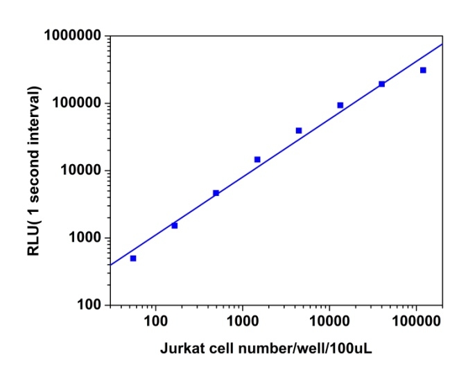
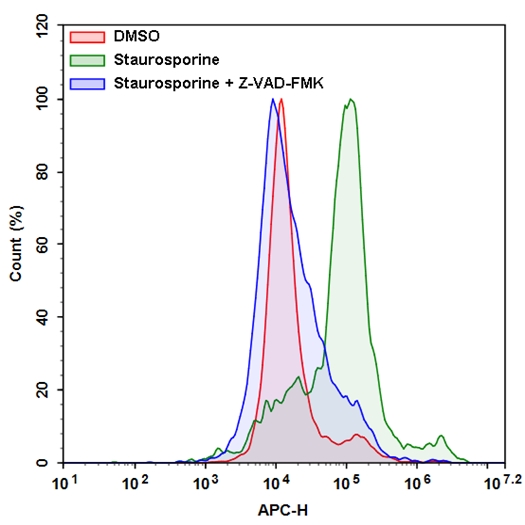
图1.在Jurkat细胞中使用Cell Meter 活细胞Caspase 8检测试剂盒对活性caspase 8进行流式细胞分析。细胞用1μM星形孢菌素处理5小时(绿色),而未处理的细胞用作对照(红色)。星形孢菌素反应被显示为蓝色的Z-VAD-FMK(胱天蛋白酶抑制剂)抑制。将细胞与iFluor 647-LETD-FMK在室温下孵育1小时。使用带有660/20 nm滤光片(APC通道)的NovoCyte流式细胞仪测量荧光强度。


参考文献

A matter of life and death for caspase 8.
Authors: Willson, Joseph
Journal: Nature reviews. Molecular cell biology (2020): 63

Cancer Cells Employ Nuclear Caspase-8 to Overcome the p53-Dependent G2/M Checkpoint through Cleavage of USP28.
Authors: Müller, Ines and Strozyk, Elwira and Schindler, Sebastian and Beissert, Stefan and Oo, Htoo Zarni and Sauter, Thomas and Lucarelli, Philippe and Raeth, Sebastian and Hausser, Angelika and Al Nakouzi, Nader and Fazli, Ladan and Gleave, Martin E and Liu, He and Simon, Hans-Uwe and Walczak, Henning and Green, Douglas R and Bartek, Jiri and Daugaard, Mads and Kulms, Dagmar
Journal: Molecular cell (2020): 970-984.e7

Caspase-8 Induces Lysosome-Associated Cell Death in Cancer Cells.
Authors: Zhong, Benfu and Liu, Miao and Bai, Changsen and Ruan, Yuxia and Wang, Yuanyuan and Qiu, Li and Hong, Yang and Wang, Xin and Li, Lifang and Li, Binghui
Journal: Molecular therapy : the journal of the American Society of Gene Therapy (2020)

Caspase-8: The double-edged sword.
Authors: Mandal, Ranadip and Barrón, Joan Compte and Kostova, Izabela and Becker, Sven and Strebhardt, Klaus
Journal: Biochimica et biophysica acta. Reviews on cancer (2020): 188357

Chrm3 protects against acinar cell necrosis by stabilizing caspase-8 expression in severe acute pancreatitis mice model.
Authors: Huang, Ning and Murtaza, Ghulam and Wang, Lujing and Luan, Jing and Wang, Xinlei and Sun, Yumiao and Wu, Xing and Tao, Yuxi and Shi, Shuoxi and Cao, Peihua and Qiao, Yu and Han, Dong and Kou, Jiayuan and Ma, Ning and Gao, Xu
Journal: Journal of cellular biochemistry (2020): 2618-2631

Cigarette smoke inhibits the NLRP3 inflammasome and leads to caspase-1 activation via the TLR4-TRIF-caspase-8 axis in human macrophages.
Authors: Buscetta, Marco and Di Vincenzo, Serena and Miele, Monica and Badami, Ester and Pace, Elisabetta and Cipollina, Chiara
Journal: FASEB journal : official publication of the Federation of American Societies for Experimental Biology (2020): 1819-1832

Dissecting DISC regulation via pharmacological targeting of caspase-8/c-FLIPL heterodimer.
Authors: Hillert, Laura K and Ivanisenko, Nikita V and Busse, Denise and Espe, Johannes and König, Corinna and Peltek, Sergey E and Kolchanov, Nikolai A and Ivanisenko, Vladimir A and Lavrik, Inna N
Journal: Cell death and differentiation (2020)

Edwardsiella piscicida type III protein EseJ suppresses apoptosis through down regulating type 1 fimbriae, which stimulate the cleavage of caspase-8.
Authors: He, Tian Tian and Zhou, Ying and Liu, Ying Li and Li, Duan You and Nie, Pin and Li, Ai Hua and Xie, Hai Xia
Journal: Cellular microbiology (2020): e13193

Expression levels of EPHB4, EFNB2 and caspase-8 are associated with clinicopathological features and progression of esophageal squamous cell cancer.
Authors: Ni, Qianzhi and Chen, Pingping and Zhu, Bing and Li, Jingjing and Xie, Dong and Ma, Xingyuan
Journal: Oncology letters (2020): 917-929

High-Concentrate Feeding to Dairy Cows Induces Apoptosis via the NOD1/Caspase-8 Pathway in Mammary Epithelial Cells.
Authors: Ul Aabdin, Zain and Cheng, Xiaoye and Dai, Hongyu and Wang, Yan and Sahito, Benazir and Roy, Animesh Chandra and Memon, Meena Arif and Shen, Xiangzhen
Journal: Genes (2020)

说明书
Cell Meter Caspase 8活性细胞结合检测试剂盒 红色荧光.pdf

PhosphoWorks 发光法ATP检测试剂盒 *温和发光* 货号21609-AAT Bioquest荧光染料

上海金畔生物科技有限公司代理AAT Bioquest荧光染料全线产品,欢迎访问AAT Bioquest荧光染料官网了解更多信息。

PhosphoWorks 发光法ATP检测试剂盒 *温和发光*

PhosphoWorks 发光法ATP检测试剂盒 *温和发光*

PhosphoWorks 发光法ATP检测试剂盒 *温和发光*    货号21609 货号 21609 存储条件 在零下15度以下保存, 避免光照
规格 1 Plate 价格 1944
Ex (nm) Em (nm)
分子量 溶剂
产品详细介绍

简要概述

PhosphoWorks 发光法ATP检测试剂盒是美国AAT Bioquest生产的用于检测ATP的试剂盒,腺苷三磷酸(ATP)在细胞能量学,代谢调节和细胞信号传导中起重要作用。 PhosphoWorks ATP检测试剂盒提供快速,简单和均匀的发光检测,用于测定哺乳动物细胞中的细胞增殖和细胞毒性。 可以以方便的96孔和384孔微量滴定板形式进行测定。 该测定的高灵敏度允许在许多生物系统,环境样品和食物中检测ATP。 这种PhosphoWorks ATP检测试剂盒具有长达4小时的稳定发光信号。 它具有稳定的发光,不需要混合或分离,并配制成具有最小的手动操作时间。金畔生物是AAT Bioquest的中国代理商,为您提供最优质的PhosphoWorks 发光法ATP检测试剂盒。 


适用仪器


发光酶标仪  
推荐孔板: 白色孔板

产品说明书

操作步骤

简要概述

1.用测试化合物(100μL/ 96孔板或25μL/ 384孔板)制备细胞(样品)
2.加入等体积的ATP工作溶液(100μL/ 96孔板或25μL/ 384孔板)
3.在室温下孵育10-20分钟
4.监测发光强度


溶液制备

1.将整瓶10 mL反应缓冲液(组分C)转移到ATP(组分B)中并充分混合。

2.将20μLATP监测酶(组分A)加入到组分B + C的瓶中并充分混合以制备ATP工作溶液。 注意:避免来自外源生物来源的潜在污染。

有关细胞样品制备的指南(点击查看)


样品试验方案

1运行ATP测定:

1.1用测试化合物处理细胞(或样品),在96孔板中加入10μL10X化合物,或在所需化合物缓冲液中加入5μL5X化合物用于384孔板。对于空白孔(没有细胞的培养基),加入相应量的化合物缓冲液。

1.2将细胞板在37℃,5%CO 2培养箱中孵育所需的一段时间,例如24,48或96小时。

1.3向每个孔中加入100μL(96孔板)或25μL(384孔板)的ATP工作溶液。

1.4在室温下孵育10-20分钟。

1.5用标准发光计监测发光强度。


2.生成标准ATP校准曲线:

        如果需要计算样品中ATP的绝对量,则应与上述分析一起生成ATP标准曲线。

2.1通过包含不含ATP的样品(作为对照)用于测量背景发光,在含有0.1%BSA的PBS缓冲液中制备一系列ATP稀释液。注意:通常ATP浓度从1 nM到10μM是合适的。

2.2将相同量的稀释的ATP溶液加入空板中(对于96孔板为100μL或对于384孔板为25μL)。

2.3加入100μL/孔(96孔板)或25μL/孔(384孔板)的ATP工作溶液。

2.4将反应混合物在室温下孵育10至20分钟。

2.5用标准光度计监测发光强度。

2.6生成ATP标准曲线。


数据分析

        从空白标准孔获得的读数(RLU)用作阴性对照。 从其他标准的读数中减去该值,以获得基线校正值。 然后,绘制标准读数以获得标准曲线和方程。 该等式可用于计算ATP样品。 我们建议使用在线线性回归计算器,该计算器可在以下位置找到:

PhosphoWorks 发光法ATP检测试剂盒 *温和发光*    货号21609

图1.使用PhosphoWorks发光ATP检测试剂盒测量ATP剂量反应。 以1秒间隔监测10μM至0.1nM的ATP浓度长达5小时。


参考文献

High throughput cell-based assay for identification of glycolate oxidase inhibitors as a potential treatment for Primary Hyperoxaluria Type 1
Authors: Mengqiao Wang, Miao Xu, Yan Long, Sonia Fargue, Noel Southall, Xin Hu, John C McKew, Christopher J Danpure, Wei Zheng
Journal: Scientific Reports (2016)

NT1014, a novel biguanide, inhibits ovarian cancer growth in vitro and in vivo
Authors: Lu Zhang, Jianjun Han, Amanda L Jackson, Leslie N Clark, Joshua Kilgore, Hui Guo, Nick Livingston, Kenneth Batchelor, Yajie Yin, Timothy P Gilliam
Journal: Journal of Hematology & Oncology (2016): 91

The Different Effects of Atorvastatin and Pravastatin on Cell Death and PARP Activity in Pancreatic NIT-1 Cells
Authors: Ya-Hui Chen, Yi-Chun Chen, Chin-San Liu, Ming-Chia Hsieh
Journal: Journal of Diabetes Research (2016)

BPA-induced DNA hypermethylation of the master mitochondrial gene PGC-1α contributes to cardiomyopathy in male rats
Authors: Ying Jiang, Wei Xia, Jie Yang, Yingshuang Zhu, Huailong Chang, Juan Liu, Wenqian Huo, Bing Xu, Xi Chen, Yuanyuan Li
Journal: Toxicology (2015): 21–31

Glutamine promotes ovarian cancer cell proliferation through the mTOR/S6 pathway
Authors: Lingqin Yuan, Xiugui Sheng, Adam K Willson, Dario R Roque, Jessica E Stine, Hui Guo, Hannah M Jones, Chunxiao Zhou, Victoria L Bae-Jump
Journal: Endocrine-related cancer (2015): 577–591

JQ1 suppresses tumor growth through downregulating LDHA in ovarian cancer
Authors: Haifeng Qiu, Amanda L Jackson, Joshua E Kilgore, Yan Zhong, Leo Li-Ying Chan, Paola A Gehrig, Chunxiao Zhou, Victoria L Bae-Jump
Journal: Oncotarget (2015): 6915

Loss of histone deacetylase Hdac1 disrupts metabolic processes in intestinal epithelial cells
Authors: Alexis Gonneaud, Naomie Turgeon, Frančois-Michel Boisvert, Frančois Boudreau, Claude Asselin
Journal: FEBS letters (2015): 2776–2783

A neutrophil intrinsic impairment affecting Rab27a and degranulation in cystic fibrosis is corrected by CFTR potentiator therapy
Authors: Kerstin Pohl, Elaine Hayes, Joanne Keenan, Michael Henry, Paula Meleady, Kevin Molloy, Bakr Jundi, David A Bergin, Cormac McCarthy, Oliver J McElvaney
Journal: Blood (2014): 999–1009

Fluorescence lifetime imaging unravels C. trachomatis metabolism and its crosstalk with the host cell
Authors: Márta Szaszák, Philipp Steven, Kensuke Shima, Regina Orzekowsky-Schroder, Gereon Huttmann, Inke R Konig, Werner Solbach, Jan Rupp
Journal: PLoS pathogens (2011): e1002108

G protein coupled receptor kinase 2 interacting protein 1 (GIT1) is a novel regulator of mitochondrial biogenesis in heart
Authors: Jinjiang Pang, Xiangbin Xu, Michael R Getman, Xi Shi, Stephen L Belmonte, Heidi Michaloski, Amy Mohan, Burns C Blaxall, Bradford C Berk
Journal: Journal of molecular and cellular cardiology (2011): 769–776


相关产品

产品名称 货号
PhosphoWorks 发光法ATP检测试剂盒* DTT – Free* Cat#21612
PhosphoWorks 发光法ATP检测试剂盒 *明亮发光* Cat#21610
PhosphoWorks 荧光ATP检测试剂盒 Cat#21620

说明书
PhosphoWorks 发光法ATP检测试剂盒 *温和发光*.pdf

巴豆醛-BSA缀合物 货号16070-AAT Bioquest荧光染料

上海金畔生物科技有限公司代理AAT Bioquest荧光染料全线产品,欢迎访问AAT Bioquest荧光染料官网了解更多信息。

巴豆醛-BSA缀合物

巴豆醛-BSA缀合物

货号 16070 存储条件 在零下15度以下保存, 避免光照
规格 100 ug 价格 3732
Ex (nm) Em (nm)
分子量 溶剂 Water
产品详细介绍

简要概述

产品基本信息

货号:16070

产品名称:巴豆醛-BSA缀合物

规格:100ug

储存条件:-15℃避光防潮

保质期:12个月


产品物理化学光谱特性

分子量:N/A

溶剂:水


产品介绍

巴豆醛是一种常见的环境污染物。烟草烟雾是巴豆醛的重要来源。巴豆醛也自然存在于某些水果和蔬菜、精选乳制品和一些酒精饮料中。除了这些外源性来源外,巴豆醛也是在脂质过氧化(LPO)过程中内生形成的,LPO可能是导致不吸烟者血清中醛类存在的一个因素。与其他不饱和醛一样,巴豆醛具有许多毒理学特性,包括在实验动物中引起多器官毒性的能力;在体外的DNA损伤和致突变性;在体内的致癌性。将巴豆醛加入牛血清白蛋白(BSA)中,制备了巴豆醛-BSA缀合物。我们的巴豆醛-牛血清白蛋白缀合物完全具有巴豆醛/蛋白质的定义比率,这是其他供应商没有提供的。它已用于制备抗巴豆醛抗体和发展巴豆醛定量分析。金畔生物是AAT Bioquest的中国代理商,为您提供最优质的巴豆醛-BSA缀合物。 


参考文献

Mercapturic Acids Derived from the Toxicants Acrolein and Crotonaldehyde in the Urine of Cigarette Smokers from Five Ethnic Groups with Differing Risks for Lung Cancer.
Authors: Park, Sungshim L and Carmella, Steven G and Chen, Menglan and Patel, Yesha and Stram, Daniel O and Haiman, Christopher A and Le Marchand, Loic and Hecht, Stephen S
Journal: PloS one (2015): e0124841

Tandem catalysis in domino olefin cross-metathesis/intramolecular oxa-conjugate cyclization: concise synthesis of 2,6-cis-substituted tetrahydropyran derivatives.
Authors: Fuwa, Haruhiko and Noguchi, Takuma and Noto, Kenkichi and Sasaki, Makoto
Journal: Organic & biomolecular chemistry (2012): 8108-12

The organocatalytic three-step total synthesis of (+)-frondosin B.
Authors: Reiter, Maud and Torssell, Staffan and Lee, Sandra and Macmillan, David W C
Journal: Chemical science (2010): 37-42

Kinetics and mechanism of protein tyrosine phosphatase 1B inactivation by acrolein.
Authors: Seiner, Derrick R and LaButti, Jason N and Gates, Kent S
Journal: Chemical research in toxicology (2007): 1315-20

Nucleophilic carbenes as organocatalysts.
Authors: Glorius, F and Hirano, K
Journal: Ernst Schering Foundation symposium proceedings (2007): 159-81

Coupling products of nucleosides with the glyoxal adduct of deoxyguanosine.
Authors: Brock, Angela K and Kozekov, Ivan D and Rizzo, Carmelo J and Harris, Thomas M
Journal: Chemical research in toxicology (2004): 1047-56

Structural and kinetic determinants of aldehyde reduction by aldose reductase.
Authors: Srivastava, S and Watowich, S J and Petrash, J M and Srivastava, S K and Bhatnagar, A
Journal: Biochemistry (1999): 42-54

Inhibition of glutathione S-transferase activity in human melanoma cells by alpha,beta-unsaturated carbonyl derivatives. Effects of acrolein, cinnamaldehyde, citral, crotonaldehyde, curcumin, ethacrynic acid, and trans-2-hexenal.
Authors: Iersel, M L and Ploemen, J P and Struik, I and van Amersfoort, C and Keyzer, A E and Schefferlie, J G and van Bladeren, P J
Journal: Chemico-biological interactions (1996): 117-32

说明书
巴豆醛-BSA缀合物.pdf

PhosphoWorks 发光法ATP检测试剂盒 *明亮发光* 货号21610-AAT Bioquest荧光染料

上海金畔生物科技有限公司代理AAT Bioquest荧光染料全线产品,欢迎访问AAT Bioquest荧光染料官网了解更多信息。

PhosphoWorks 发光法ATP检测试剂盒 *明亮发光*

PhosphoWorks 发光法ATP检测试剂盒 *明亮发光*

PhosphoWorks 发光法ATP检测试剂盒 *明亮发光*    货号21610 货号 21610 存储条件 在零下15度以下保存, 避免光照
规格 1 Plate 价格 1944
Ex (nm) Em (nm)
分子量 溶剂
产品详细介绍

简要概述

PhosphoWorks 发光法ATP检测试剂盒 *明亮发光*是美国AAT Bioquest生产的用于检测ATP的试剂盒,三磷酸腺苷(ATP)在细胞能量生成,代谢调节和细胞信号传导中起着基本作用。 PhosphoWorks ATP检测试剂盒提供了一种快速,简单且均一的发光检测方法,用于测定哺乳动物细胞中的细胞增殖和细胞毒性。 该测定可以以方便的96孔和384孔微量滴定板形式进行。 此测定法的高灵敏度允许在许多生物系统,环境样品和食品中检测ATP。 该PhosphoWorks ATP分析试剂盒每孔可检测低至10个细胞。 它具有稳定的发光,无需混合或分离,并且配方具有最小的动手时间。金畔生物是AAT Bioquest的中国代理商,为您提供最优质的PhosphoWorks 发光法ATP检测试剂盒 *明亮发光*。 


适用仪器


发光酶标仪  
推荐孔板: 白色孔板

产品说明书

样品实验方案

简要概述

1.用测试化合物(100 µL / 96孔板或25 µL / 384孔板)制备细胞(样品)
2.加入等体积的ATP工作溶液(100 µL / 96孔板或25 µL / 384孔板)
3.在室温下孵育10-20分钟
4.监控发光强度


溶液配制

工作溶液配制

1.将全部反应缓冲液(组分C,10 mL)转移到ATP传感器(组分B)中,并充分混合。

2.将20 µL ATP监测酶(组分A)添加到组分B + C的瓶子中,并充分混合以制成ATP工作溶液。 注意:避免来自外源性生物来源的潜在ATP污染。

点击查看细胞制备方案


样品操作

1.运行ATP分析:

1.1通过在所需化合物缓冲液中加入10 µL的10X化合物(用于96孔板)或5 µL的5X化合物(用于384孔板)来用测试化合物处理细胞(或样品)。对于空白孔(没有细胞的培养基),添加相应量的化合物缓冲液。

1.2将细胞板在37°C,5%CO2培养箱中孵育所需的时间,例如24、48或96小时。

1.3向每个孔中添加100 µL(96孔板)或25 µL(384孔板)的ATP工作溶液。

1.4在室温下孵育10-20分钟。

1.5使用标准发光计监控发光强度。

2.生成标准ATP校准曲线:

如果需要计算样品中ATP的绝对量,则应与上述测定法一起生成ATP标准曲线。

2.1通过包含不含ATP的样品(作为对照)来测量背景发光,在含0.1%BSA的PBS缓冲液中稀释一系列ATP。注意:通常,ATP浓度从1 nM到10 µM是合适的。

2.2将相同量的稀释的ATP溶液添加到一个空板中(对于96孔板是100 µL,对于384孔板是25 µL)。

2.3加入100 µL /孔(96孔板)或25 µL /孔(384孔板)的ATP工作溶液。

2.4将反应混合物在室温下孵育10至20分钟。

2.5使用标准发光计监控发光强度。

2.6生成ATP标准曲线。


参考文献

Hepatitis B Surface Antigen Activates Unfolded Protein Response in Forming Ground Glass Hepatocytes of Chronic Hepatitis B
Authors: Yao Li, Yuchen Xia, Xiaoming Cheng, David E Kleiner, Stephen M Hewitt, Julia Sproch, Tong Li, Hui Zhuang, T Jake Liang
Journal: Viruses (2019): 386

High throughput cell-based assay for identification of glycolate oxidase inhibitors as a potential treatment for Primary Hyperoxaluria Type 1
Authors: Mengqiao Wang, Miao Xu, Yan Long, Sonia Fargue, Noel Southall, Xin Hu, John C McKew, Christopher J Danpure, Wei Zheng
Journal: Scientific Reports (2016)

NT1014, a novel biguanide, inhibits ovarian cancer growth in vitro and in vivo
Authors: Lu Zhang, Jianjun Han, Amanda L Jackson, Leslie N Clark, Joshua Kilgore, Hui Guo, Nick Livingston, Kenneth Batchelor, Yajie Yin, Timothy P Gilliam
Journal: Journal of Hematology & Oncology (2016): 91

The Different Effects of Atorvastatin and Pravastatin on Cell Death and PARP Activity in Pancreatic NIT-1 Cells
Authors: Ya-Hui Chen, Yi-Chun Chen, Chin-San Liu, Ming-Chia Hsieh
Journal: Journal of Diabetes Research (2016)

BPA-induced DNA hypermethylation of the master mitochondrial gene PGC-1α contributes to cardiomyopathy in male rats
Authors: Ying Jiang, Wei Xia, Jie Yang, Yingshuang Zhu, Huailong Chang, Juan Liu, Wenqian Huo, Bing Xu, Xi Chen, Yuanyuan Li
Journal: Toxicology (2015): 21–31

Glutamine promotes ovarian cancer cell proliferation through the mTOR/S6 pathway
Authors: Lingqin Yuan, Xiugui Sheng, Adam K Willson, Dario R Roque, Jessica E Stine, Hui Guo, Hannah M Jones, Chunxiao Zhou, Victoria L Bae-Jump
Journal: Endocrine-related cancer (2015): 577–591

JQ1 suppresses tumor growth through downregulating LDHA in ovarian cancer
Authors: Haifeng Qiu, Amanda L Jackson, Joshua E Kilgore, Yan Zhong, Leo Li-Ying Chan, Paola A Gehrig, Chunxiao Zhou, Victoria L Bae-Jump
Journal: Oncotarget (2015): 6915

Loss of histone deacetylase Hdac1 disrupts metabolic processes in intestinal epithelial cells
Authors: Alexis Gonneaud, Naomie Turgeon, Frančois-Michel Boisvert, Frančois Boudreau, Claude Asselin
Journal: FEBS letters (2015): 2776–2783

A neutrophil intrinsic impairment affecting Rab27a and degranulation in cystic fibrosis is corrected by CFTR potentiator therapy
Authors: Kerstin Pohl, Elaine Hayes, Joanne Keenan, Michael Henry, Paula Meleady, Kevin Molloy, Bakr Jundi, David A Bergin, Cormac McCarthy, Oliver J McElvaney
Journal: Blood (2014): 999–1009

Fluorescence lifetime imaging unravels C. trachomatis metabolism and its crosstalk with the host cell
Authors: Márta Szaszák, Philipp Steven, Kensuke Shima, Regina Orzekowsky-Schroder, Gereon Huttmann, Inke R Konig, Werner Solbach, Jan Rupp
Journal: PLoS pathogens (2011): e1002108


相关产品

产品名称 货号
PhosphoWorks 发光法ATP检测试剂盒 *温和发光* Cat#21609
PhosphoWorks 发光法ATP检测试剂盒* DTT – Free* Cat#21612
PhosphoWorks 比色ATP分析试剂盒 Cat#21617

说明书
PhosphoWorks 发光法ATP检测试剂盒 *明亮发光*.pdf

巴豆醛-HSA(人血清白蛋白)缀合物 货号16072-AAT Bioquest荧光染料

上海金畔生物科技有限公司代理AAT Bioquest荧光染料全线产品,欢迎访问AAT Bioquest荧光染料官网了解更多信息。

巴豆醛-HSA(人血清白蛋白)缀合物

巴豆醛-HSA(人血清白蛋白)缀合物

货号 16072 存储条件 在零下15度以下保存, 避免光照
规格 100 ug 价格 4992
Ex (nm) Em (nm)
分子量 溶剂 Water
产品详细介绍

简要概述

产品基本信息

货号:16072

产品名称:巴豆醛-HSA(人血清白蛋白)缀合物

规格:100ug

储存条件:-15℃避光防潮

保质期:12个月


产品物理化学光谱特性

分子量:N/A

溶剂:水


产品介绍

巴豆醛是一种常见的环境污染物。烟草烟雾是巴豆醛的重要来源。巴豆醛也自然存在于某些水果和蔬菜、精选乳制品和一些酒精饮料中。除了这些外源性来源外,巴豆醛也是在脂质过氧化(LPO)过程中内生形成的,LPO可能是导致不吸烟者血清中醛类存在的一个因素。与其他不饱和醛一样,巴豆醛具有许多毒理学特性,包括在实验动物中引起多器官毒性的能力;在体外的DNA损伤和致突变性;在体内的致癌性。将巴豆醛加入牛血清白蛋白(BSA)中,制备了巴豆醛-BSA缀合物。我们的巴豆醛-牛血清白蛋白缀合物完全具有巴豆醛/蛋白质的定义比率,这是其他供应商没有提供的。它已用于制备抗巴豆醛抗体和发展巴豆醛定量分析。金畔生物是AAT Bioquest的中国代理商,为您提供最优质的巴豆醛-HSA(人血清白蛋白)缀合物。 


参考文献

Crotonaldehyde-induced alterations in testicular enzyme function and hormone levels, and apoptosis in the testes of male Wistar rats are associated with oxidative damage.
Authors: Zhang, Biao and Wei, Ping and Men, Jinlong and Zhang, Shuman and Shao, Hua and Zhang, Zhihu
Journal: Toxicology mechanisms and methods (2020): 19-32

Resolution and Quantitation of Mercapturic Acids Derived from Crotonaldehyde, Methacrolein, and Methyl Vinyl Ketone in the Urine of Smokers and Nonsmokers.
Authors: Chen, Menglan and Carmella, Steven G and Li, Yupeng and Zhao, Yingchun and Hecht, Stephen S
Journal: Chemical research in toxicology (2020): 669-677

A computational foray into the mechanism and catalysis of the adduct formation reaction of guanine with crotonaldehyde.
Authors: Kroeger, Asja A and Karton, Amir
Journal: Journal of computational chemistry (2019): 630-637

Autophagy in Crotonaldehyde-Induced Endothelial Toxicity.
Authors: Lee, Seung Eun and Park, Hye Rim and Park, Cheung-Seog and Ahn, Hyun-Jong and Cho, Jeong-Je and Lee, Jongsung and Park, Yong Seek
Journal: Molecules (Basel, Switzerland) (2019)

Autophagy induced by low concentrations of crotonaldehyde promotes apoptosis and inhibits necrosis in human bronchial epithelial cells.
Authors: Wang, Limeng and Li, Xiang and Yang, Zhihua and Zhu, Maoxiang and Xie, Jianping
Journal: Ecotoxicology and environmental safety (2019): 169-177

Effect of influent pH on hydrolytic acidification performance and bacterial community structure in EGSB for pretreating crotonaldehyde manufacture wastewater after ozonation.
Authors: Liu, Tao and Shen, Zhiqiang and Zhang, Chunyu and Song, Yudong and Li, Jie and Yang, Zongpu and Song, Guangqing and Han, Zhenfeng and Zhou, Yuexi
Journal: Water science and technology : a journal of the International Association on Water Pollution Research (2019): 1174-1183

Long-term exposure to crotonaldehyde causes heart and kidney dysfunction through induction of inflammatory and oxidative damage in male Wistar rats.
Authors: Zhang, Biao and Li, Shuangshuang and Men, Jinlong and Peng, Cheng and Shao, Hua and Zhang, Zhihu
Journal: Toxicology mechanisms and methods (2019): 263-275

Pocketlike Active Site of Rh1/MoS2 Single-Atom Catalyst for Selective Crotonaldehyde Hydrogenation.
Authors: Lou, Yang and Zheng, Yongping and Li, Xu and Ta, Na and Xu, Jia and Nie, Yifan and Cho, Kyeongjae and Liu, Jingyue
Journal: Journal of the American Chemical Society (2019): 19289-19295

Toxic trans-crotonaldehyde in mitochondria intercepted by oxyresveratrol contributing to anticancer.
Authors: Su, Yanbin and Sun, Chengyu and Chen, Yan and Liu, Shichang and Jing, Ning and Li, Shuxin
Journal: IUBMB life (2019): 1014-1020

[Study on lung injury induced by subchronic exposure to crotonaldehyde in male rats].
Authors: Li, S S and Zhang, B and Zhang, S M and Zhang, Z H and Wei, Y N
Journal: Zhonghua lao dong wei sheng zhi ye bing za zhi = Zhonghua laodong weisheng zhiyebing zazhi = Chinese journal of industrial hygiene and occupational diseases (2019): 728-731

说明书
巴豆醛-HSA(人血清白蛋白)缀合物.pdf

PhosphoWorks 发光法ATP检测试剂盒* DTT – Free* 货号21612-AAT Bioquest荧光染料

上海金畔生物科技有限公司代理AAT Bioquest荧光染料全线产品,欢迎访问AAT Bioquest荧光染料官网了解更多信息。

PhosphoWorks 发光法ATP检测试剂盒* DTT – Free*

PhosphoWorks 发光法ATP检测试剂盒* DTT – Free*

PhosphoWorks 发光法ATP检测试剂盒* DTT - Free*    货号21612 货号 21612 存储条件 在零下15度以下保存, 避免光照
规格 1 Plate 价格 1944
Ex (nm) Em (nm)
分子量 溶剂
产品详细介绍

简要概述

PhosphoWorks 发光法ATP检测试剂盒是美国AAT Bioquest生产的用于检测ATP的试剂盒,腺苷三磷酸(ATP)在细胞能量学,代谢调节和细胞信号传导中起重要作用。 PhosphoWorks ATP检测试剂盒提供快速,简单和均匀的发光检测,用于测定哺乳动物细胞中的细胞增殖和细胞毒性。 可以以方便的96孔和384孔微量滴定板形式进行测定。 该测定的高灵敏度允许在许多生物系统,环境样品和食物中检测ATP。 该PhosphoWorks ATP检测试剂盒不使用DTT,并且具有长达4小时的稳定发光信号。 它具有稳定的发光,不需要混合或分离,并配制成具有最小的手动操作时间。金畔生物是AAT Bioquest的中国代理商,为您提供最优质的PhosphoWorks 发光法ATP检测试剂盒。 


适用仪器


发光酶标仪  
推荐孔板: 白色孔板

产品说明书

操作步骤

简要概述

1.用测试化合物(100μL/ 96孔板或25μL/ 384孔板)制备细胞(样品)
2.加入等体积的ATP工作溶液(100μL/ 96孔板或25μL/ 384孔板)
3.在室温下孵育10-20分钟
4.监测发光强度


制备工作溶液

1.将10 mL反应缓冲液(组分C)转移到ATP传感器(组分B)中并充分混合。

2.将20μLATP监测酶(组分A)加入到组分B + C的瓶中并充分混合以制备ATP工作溶液。 注意:避免来自外源生物来源的潜在ATP污染。

有关细胞样品制备的指南(点击查看)


样品实验方案

1.运行ATP测定:

1.1用测试化合物处理细胞(或样品),在96孔板中加入10μL10X化合物,或在所需化合物缓冲液中加入5μL5X化合物用于384孔板。对于空白孔(没有细胞的培养基),加入相应量的化合物缓冲液。

1.2将细胞板在37℃,5%CO 2培养箱中孵育所需的一段时间,例如24,48或96小时。

1.3向每个孔中加入100μL(96孔板)或25μL(384孔板)的ATP工作溶液。

1.4在室温下孵育10-20分钟。

1.5用标准发光计监测发光强度。


2.生成标准ATP校准曲线:

        如果需要计算样品中ATP的绝对量,则应与上述分析一起生成ATP标准曲线。

2.1通过包含不含ATP的样品(作为对照)在含有0.1%BSA的PBS缓冲液中制备ATP系列稀释液以测量背景发光。注意:通常ATP浓度范围为0.1 nM至1μM是合适的。

2.2将相同量的稀释的ATP溶液加入空板中(对于96孔板为100μL或对于384孔板为25μL)。

2.3加入100μL/孔(96孔板)或25μL/孔(384孔板)的ATP工作溶液。

2.4将反应混合物在室温下孵育10至20分钟。

2.5用标准发光计记录发光强度。

2.6生成ATP标准曲线。


数据分析

        从空白标准孔获得的读数(RLU)用作阴性对照。 从其他标准的读数中减去该值,以获得基线校正值。 然后,绘制标准读数以获得标准曲线和方程。 该等式可用于计算ATP样品。 我们建议使用在线线性回归计算器,该计算器可在以下位置找到:

PhosphoWorks 发光法ATP检测试剂盒* DTT - Free*    货号21612

图1.使用NOVOstar板读数器(BMG Labtech)在96孔白板上用PhosphoWorks TM发光ATP测定试剂盒* DTT-Free *测量ATP剂量响应。 该试剂盒可在20分钟内检测到(3 pmole /孔)0.03 nM ATP。 积分时间为1秒。 半衰期超过2小时。


参考文献

High throughput cell-based assay for identification of glycolate oxidase inhibitors as a potential treatment for Primary Hyperoxaluria Type 1
Authors: Mengqiao Wang, Miao Xu, Yan Long, Sonia Fargue, Noel Southall, Xin Hu, John C McKew, Christopher J Danpure, Wei Zheng
Journal: Scientific Reports (2016)

NT1014, a novel biguanide, inhibits ovarian cancer growth in vitro and in vivo
Authors: Lu Zhang, Jianjun Han, Amanda L Jackson, Leslie N Clark, Joshua Kilgore, Hui Guo, Nick Livingston, Kenneth Batchelor, Yajie Yin, Timothy P Gilliam
Journal: Journal of Hematology & Oncology (2016): 91

The Different Effects of Atorvastatin and Pravastatin on Cell Death and PARP Activity in Pancreatic NIT-1 Cells
Authors: Ya-Hui Chen, Yi-Chun Chen, Chin-San Liu, Ming-Chia Hsieh
Journal: Journal of Diabetes Research (2016)

BPA-induced DNA hypermethylation of the master mitochondrial gene PGC-1α contributes to cardiomyopathy in male rats
Authors: Ying Jiang, Wei Xia, Jie Yang, Yingshuang Zhu, Huailong Chang, Juan Liu, Wenqian Huo, Bing Xu, Xi Chen, Yuanyuan Li
Journal: Toxicology (2015): 21–31

Glutamine promotes ovarian cancer cell proliferation through the mTOR/S6 pathway
Authors: Lingqin Yuan, Xiugui Sheng, Adam K Willson, Dario R Roque, Jessica E Stine, Hui Guo, Hannah M Jones, Chunxiao Zhou, Victoria L Bae-Jump
Journal: Endocrine-related cancer (2015): 577–591

JQ1 suppresses tumor growth through downregulating LDHA in ovarian cancer
Authors: Haifeng Qiu, Amanda L Jackson, Joshua E Kilgore, Yan Zhong, Leo Li-Ying Chan, Paola A Gehrig, Chunxiao Zhou, Victoria L Bae-Jump
Journal: Oncotarget (2015): 6915

Loss of histone deacetylase Hdac1 disrupts metabolic processes in intestinal epithelial cells
Authors: Alexis Gonneaud, Naomie Turgeon, Frančois-Michel Boisvert, Frančois Boudreau, Claude Asselin
Journal: FEBS letters (2015): 2776–2783

A neutrophil intrinsic impairment affecting Rab27a and degranulation in cystic fibrosis is corrected by CFTR potentiator therapy
Authors: Kerstin Pohl, Elaine Hayes, Joanne Keenan, Michael Henry, Paula Meleady, Kevin Molloy, Bakr Jundi, David A Bergin, Cormac McCarthy, Oliver J McElvaney
Journal: Blood (2014): 999–1009

Fluorescence lifetime imaging unravels C. trachomatis metabolism and its crosstalk with the host cell
Authors: Márta Szaszák, Philipp Steven, Kensuke Shima, Regina Orzekowsky-Schroder, Gereon Huttmann, Inke R Konig, Werner Solbach, Jan Rupp
Journal: PLoS pathogens (2011): e1002108

G protein coupled receptor kinase 2 interacting protein 1 (GIT1) is a novel regulator of mitochondrial biogenesis in heart
Authors: Jinjiang Pang, Xiangbin Xu, Michael R Getman, Xi Shi, Stephen L Belmonte, Heidi Michaloski, Amy Mohan, Burns C Blaxall, Bradford C Berk
Journal: Journal of molecular and cellular cardiology (2011): 769–776


相关产品

产品名称 货号
PhosphoWorks 发光法ATP检测试剂盒 *温和发光* Cat#21609
PhosphoWorks 发光法ATP检测试剂盒 *明亮发光* Cat#21610
PhosphoWorks 荧光ATP检测试剂盒 Cat#21620

说明书
PhosphoWorks 发光法ATP检测试剂盒* DTT – Free*.pdf

VIC亚磷酰胺(停产) 货号6080-AAT Bioquest荧光染料

上海金畔生物科技有限公司代理AAT Bioquest荧光染料全线产品,欢迎访问AAT Bioquest荧光染料官网了解更多信息。

VIC亚磷酰胺(停产)

VIC亚磷酰胺(停产)

VIC亚磷酰胺(停产)    货号6080 货号 6080 存储条件 在零下15度以下保存, 避免光照
规格 50 umoles 价格 0
Ex (nm) 526 Em (nm) 543
分子量 1023.38 溶剂 MeCN
产品详细介绍

简要概述

产品基本信息

货号:6080

产品名称:VIC亚磷酰胺

规格:50umoles

储存条件:-15℃避光防潮

保质期:12个月


产品物理化学光谱特性

分子量:1023.38

溶剂:MeCN


产品介绍

VIC是一种荧光染料,最初由Applied Biosystems(现为Thermofisher)开发。VIC的光谱为可见光谱的绿黄色部分。它用于在5’末端荧光标记寡核苷酸(用作各种实时PCR,杂交和基于荧光的遗传分析应用中的探针)。VIC亚磷酰胺是将VIC标签掺入寡核苷酸中最方便的产品。金畔生物是AAT Bioquest的中国代理商,为您提供最优质的VIC亚磷酰胺。


参考文献

Optimization of digital droplet polymerase chain reaction for quantification of genetically modified organisms.
Authors: Gerdes, Lars and Iwobi, Azuka and Busch, Ulrich and Pecoraro, Sven
Journal: Biomolecular detection and quantification (2016): 9-20

Simultaneous detection of the main black aspergilli responsible for ochratoxin A (OTA) contamination in grapes by multiplex real-time polymerase chain reaction.
Authors: Selma, M V and Martínez-Culebras, P V and Elizaquível, P and Aznar, R
Journal: Food additives & contaminants. Part A, Chemistry, analysis, control, exposure & risk assessment (2009): 180-8

Multiplex real-time PCR for the detection and differentiation of equid herpesvirus 1 (EHV-1) and equid herpesvirus 4 (EHV-4).
Authors: Diallo, Ibrahim S and Hewitson, Glen and Wright, Lucia L and Kelly, Mark A and Rodwell, Barry J and Corney, Bruce G
Journal: Veterinary microbiology (2007): 93-103

Detection and quantitation of akabane and aino viruses by multiplex real-time reverse-transcriptase PCR.
Authors: Stram, Yehuda and Kuznetzova, Larisa and Guini, Merisol and Rogel, Arie and Meirom, Ruth and Chai, Dalia and Yadin, Hagai and Brenner, Jackob
Journal: Journal of virological methods (2004): 147-54

Structure of the cytochrome c oxidase complex of rat liver. 2. Topological orientation of polypeptides in the membrane as studied by proteolytic digestion and immunoblotting.
Authors: Jarausch, J and Kadenbach, B
Journal: European journal of biochemistry (1985): 219-25

说明书
VIC亚磷酰胺(停产).pdf

红色荧光示踪探针 CytoTrace CFDA 货号22016-AAT Bioquest荧光染料

上海金畔生物科技有限公司代理AAT Bioquest荧光染料全线产品,欢迎访问AAT Bioquest荧光染料官网了解更多信息。

红色荧光示踪探针 CytoTrace CFDA

红色荧光示踪探针 CytoTrace CFDA

红色荧光示踪探针 CytoTrace CFDA    货号22016 货号 22016 存储条件 在零下15度以下保存, 避免光照
规格 1 mg 价格 2604
Ex (nm) 562 Em (nm) 576
分子量 652.43 溶剂 DMSO
产品详细介绍

简要概述

CytoTrace 红色荧光探针保留了Cy3 / TRITC的光谱特性,因此可以使用荧光显微镜或流式细胞仪轻松检测其信号。它自由地穿过细胞膜,并在与细胞成分反应后转化为不渗透细胞的产物。细胞不渗透的反应产物经过数代传给子细胞,但没有转移到群体中的相邻细胞。装有CytoTrace Red探针的细胞通常可以发出荧光,并且至少可以存活24小时,这使该探针成为了出色的长期细胞示踪剂。染色图案可以用甲醛或戊二醛固定,用于信号放大和其他应用。它的光谱与GFP或FITC标记的抗体的光谱很好分离。

点击查看光谱

产品说明书

操作步骤

该协议仅提供指南,应根据您的特定需求进行修改。

1.准备2-10 mMDMSO储备溶液

对于#22014添加45微升DMSO成50 μ 克小瓶使2mM的储备溶液(1毫克/毫升,相当于1.8毫摩尔);

对于#22015添加36微升DMSO成50 μ 克小瓶制成10mM储备溶液(1毫克/毫升,相当于1.46毫摩尔);

对于#22016,添加153 uL ml DMSO以制成10 mM储备溶液(1 mg / ml相当于1.53 mM);

对于#22017,添加215μLDMSO以制成10 mM储备溶液(1 mg / ml相当于2.15 mM);

对于#22020,将4.2 mg溶于1 ml DMSO中制成10 mM储备溶液(1 mg / ml相当于〜2.4 mM);

注意:储备溶液应及时使用;应将所有剩余的溶液等分并在< -20 o C下冷冻。避免重复冻融循环,并避光

2.准备染料工作液

在使用前,通过用Hanks和20 mM Hepes缓冲液(HHBS)或您选择的pH 7缓冲液稀释步骤1中的DMSO储备溶液,准备好1至20 µM的染料工作溶液。

3.用流式细胞仪或荧光显微镜分析细胞:

3.1用测试化合物处理细胞所需的时间。

3.2离心细胞,每管得到2-10 x105细胞。

3.3将细胞重悬于500 µL 的染料工作溶液中(来自步骤2)。

3.4将细胞与染料溶液在室温或37° C下避光放置15至30分钟。

3.5从细胞中除去染料工作溶液,用HHBS或您选择的缓冲液洗涤细胞。将细胞重悬于500 µL预热的HHBS或培养基中,每管可得到2-10 x 10 5个细胞。

3.6用流式细胞仪(FL1通道) 或荧光显微镜监测Ex / Em = 490/520 nm处的荧光变化。

注意:对于细菌细胞染色:将母液在通过测试细菌过夜生长而预先调节的营养肉汤中以1:800的比例稀释时,染色是最有效的,但是也可以使用新鲜的营养肉汤或PBS。细菌悬浮液应用PBS稀释至105 – 10 7每毫升。将1 ml溶液加到.45 µm过滤器(25mm)上并真空过滤除去溶液,然后加入1ml染料溶液并在室温下孵育5-10分钟,即可对细菌染色。


参考文献

Fluorescence-Based Transport Assays Revisited in a Human Renal Proximal Tubule Cell Line
Authors: Caetano-Pinto P, Janssen MJ, Gijzen L, Verscheijden L, Wilmer MJ, Masereeuw R.
Journal: Mol Pharm (2016): 933

The variable chemotherapeutic response of Malabaricone-A in leukemic and solid tumor cell lines depends on the degree of redox imbalance
Authors: Manna A, De Sarkar S, De S, Bauri AK, Chattopadhyay S, Chatterjee M.
Journal: Phytomedicine (2015): 713

Cell membrane tracker based on restriction of intramolecular rotation
Authors: Zhang C, Jin S, Yang K, Xue X, Li Z, Jiang Y, Chen WQ, Dai L, Zou G, Liang XJ.
Journal: ACS Appl Mater Interfaces (2014): 8971

A multiple model probability hypothesis density tracker for time-lapse cell microscopy sequences
Authors: Rezatofighi SH, Gould S, Vo BN, Mele K, Hughes WE, Hartley R.
Journal: Inf Process Med Imaging (2013): 110

Evaluation of stability and sensitivity of cell fluorescent labels when used for cell migration
Authors: Beem E, Segal MS.
Journal: J Fluoresc (2013): 975

TLM-Tracker: software for cell segmentation, tracking and lineage analysis in time-lapse microscopy movies
Authors: Klein J, Leupold S, Biegler I, Biedendieck R, Munch R, Jahn D.
Journal: Bioinformatics (2012): 2276

Horizontal DNA transfer from donor to host cells as an alternative mechanism of epithelial chimerism after allogeneic hematopoietic cell transplantation
Authors: Waterhouse M, Themeli M, Bertz H, Zoumbos N, Finke J, Spyridonidis A.
Journal: Biol Blood Marrow Transplant (2011): 319

The exocytosis of fluorescent nanodiamond and its use as a long-term cell tracker
Authors: Fang CY, Vaijayanthimala V, Cheng CA, Yeh SH, Chang CF, Li CL, Chang HC.
Journal: Small (2011): 3363

The interplay between Leishmania promastigotes and human Natural Killer cells in vitro leads to direct lysis of Leishmania by NK cells and modulation of NK cell activity by Leishmania promastigotes
Authors: Lieke T, Nylen S, Eidsmo L, Schmetz C, Berg L, Akuffo H.
Journal: Parasitology (2011): 1898

Cell electrofusion visualized with fluorescence microscopy
Authors: Trontelj K, Usaj M, Miklavcic D.
Journal: J Vis Exp. (2010)

说明书
红色荧光示踪探针 CytoTrace CFDA.pdf